Perfusion computed tomography allows studying cerebral hemodynamics at the level of capillaries.
Perfusion computed tomography is the assessment of the brain parenchyma, its blood supply, and in case of the impairment, determines the level of brain matter change.
During the acute ischemic stroke, perfusion is an essential study, as it differentiates dead and susceptible cells (tissues) due to ischemia. Identifying these changes (differentiation) are important for the proper management of the patient. The method significantly reduces the cases of disability and death.
From the first minutes of the disease, this method can detect the pathological process, the area of ischemia and the determine the penumbra (reduced or marginal zone). The color map allows us to detect even the smallest areas of damage.
The National Center of Surgery is equipped with multislice computed tomography Toshiba Aquilon RXL. The CT machine is a leader in its class, to which an additional function – perfusion has been added.
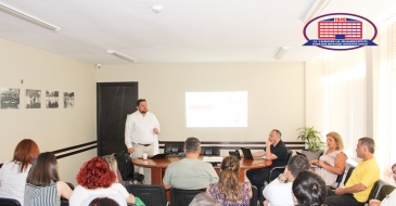
The radiologist at the National Center of Surgery Erekle Gigadze talks about the perfusion.
“During ischemic stroke, if perfusion detects cells at high risk of death, so-called penumbra cells, antithrombotic treatment is performed - thrombolysis and (or) thrombectomy. Tissues under risk return to the original function after the procedure, ie they are saved. If penumbra area is not found, then the above-mentioned methods of treatment will have no effect and they can also cause harm. That is why patients having an ischemic stroke, especially during the first hours, need to have a CT scan with perfusion immediately "- says Mr. Erekle.
This procedure with standard contrast is identical to the computed tomography, but in this case, the program installed in the device gives the radiologist images of different colors. The received tomograms are assessed by the radiologist.
The most important research that saves patients with stroke not only from disability, but also saves a life, takes only 15-20 minutes with reconstructions and evaluations.
Perfusion computed tomography is successfully used in the following cases:
- Blood flow disruption during the ischemic stroke;
- Blood flow disturbances in traumatic brain injuries;
- Assessment of hemodynamics in brain tumors;
- Detection of occlusive diseases, chronic stenosis of the intra- and extracranial arteries, hidden ischemic processes in the brain parenchyma due to critical stenosis of the carotid artery;
- Determination of the exact area for biopsy in brain tumors;
- Monitoring of a brain tumor after radiation and chemotherapy.
The National Center of Surgery offers a promotion, Perfusion computed tomography.
If you want to know more about the promotion, see the details!
National Center of Surgery address – Tbilisi, Digomi Chachava street N5
You can contact the Call-center of the National Center of Surgery at 577 119119 or 2 02 25 25;
Wish you health!