Good to know (MRI)
Magnetic resonance imaging, commonly referred to as MRI/MRT, is a safe and efficient imaging technique used in radiology for the study of the body's anatomy and physiology.
The device creates images by using radio waves and a magnetic field. The study is safe for children and pregnant women starting in the second trimester since no ionizing radiation is utilized and no human tissues or internal organs are exposed to radiation;
What is meant by 1.5T; 3.0 T? - This shows the device's power; the more powerful the information gathered throughout the study, the higher the Tesla rating.
The following tools are available at the National Center of Surgery:
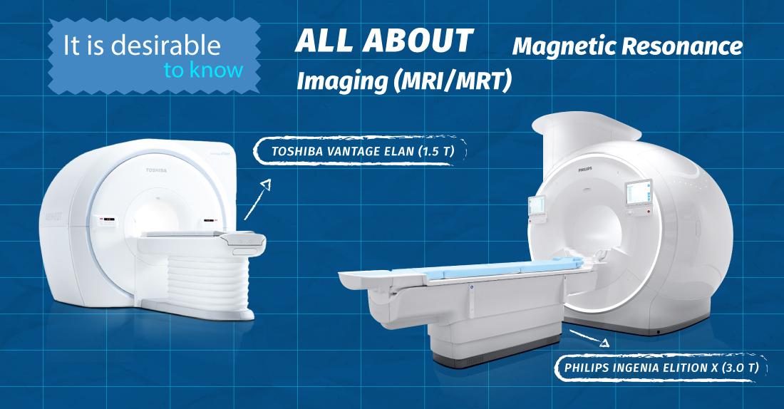
- Contemporary tomographs:
- One of the greatest models on the medical market is the Toshiba Vantage Elan 1.5 Tesla, with its features and the newest technical packages.
- 3.0 Tesla, digital MRI (PHILIPS INGENIA ELITION X) - its uniqueness is due to artificial intelligence and the latest technical capabilities, which are focused on patient comfort and provide the doctor with the most accurate image, without artifacts (false images)!
- The absence of claustrophobia, the sensation of open space, and the largest tunnel;
- Video and audio services - 3D cinema;
- Practically unlimited patient weight - up to 250 kg;
- Anatomical bed area adjusted to the patient's spine;
- Half the examination time!
- With MRI (MRT/MRI) magnetic resonance imaging it is possible to:
Magnetic resonance imaging (MRI) can:
- Detailed structural and functional analysis of the brain and diagnosis of pathological processes at the initial stage;
- Magnetic resonance angiography of the main blood vessels of the brain and neck - analysis of the main blood supply of the brain and neck;
- Detailed evaluation of the spine and spinal cord;
- Magnetic resonance angiography, dynamic evaluation of the kidney, non-contrast magnetic resonance cholangiopancreatography of the biliary tract, and assessment of the abdominal organs, including the liver, spleen, pancreas, adrenal glands, and kidneys;
- Early illness diagnosis using magnetic resonance imaging of pelvic organ diseases;
- Musculoskeletal system structural and functional analysis (any joint, soft tissues).
What is a contrast study? - A specific method of research. A contrast agent is injected into a vein, against the background of which blood vessels and various formations are more clearly visible. Research with intravenous contrast is not routinely performed. . It is prescribed when more detail is needed in a suspicious area. It is used in the case of CT, MRI studies. MRI studies are performed every day, including weekends, 24/7.
Make an appointment at your preferred time: +995 577 119 119 or +995 322 02 25 25
Address: Tbilisi, Dighomi Chachava №7
Wish you health!
Give us a call