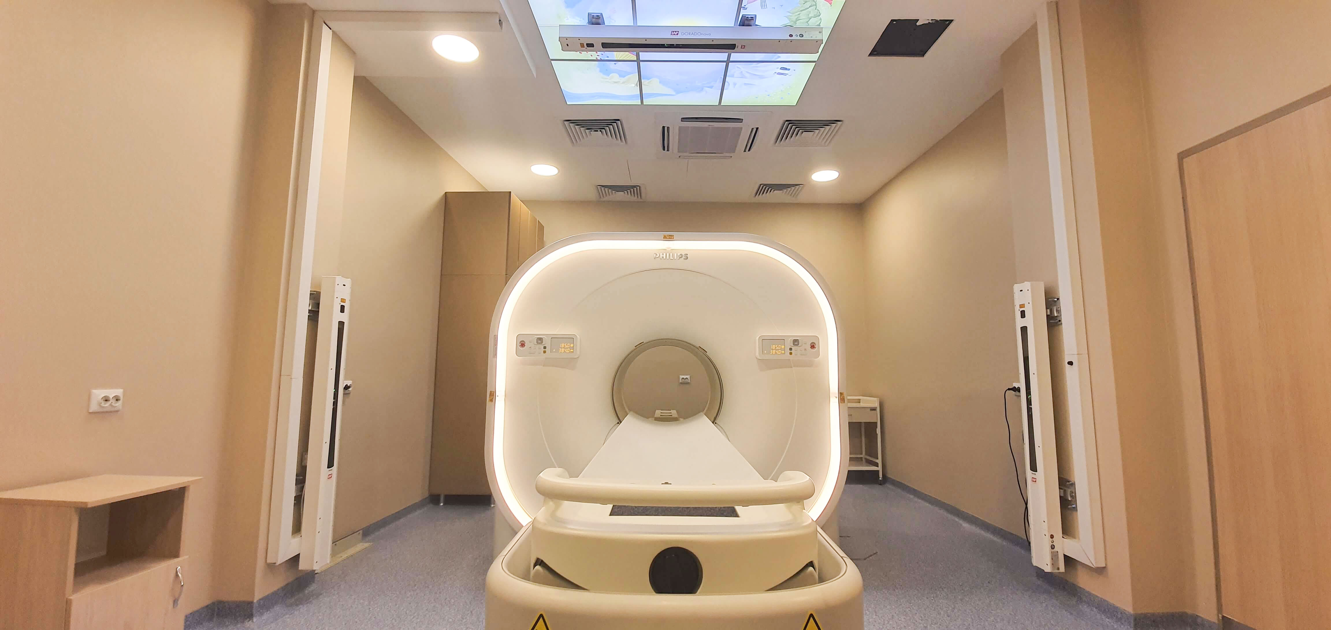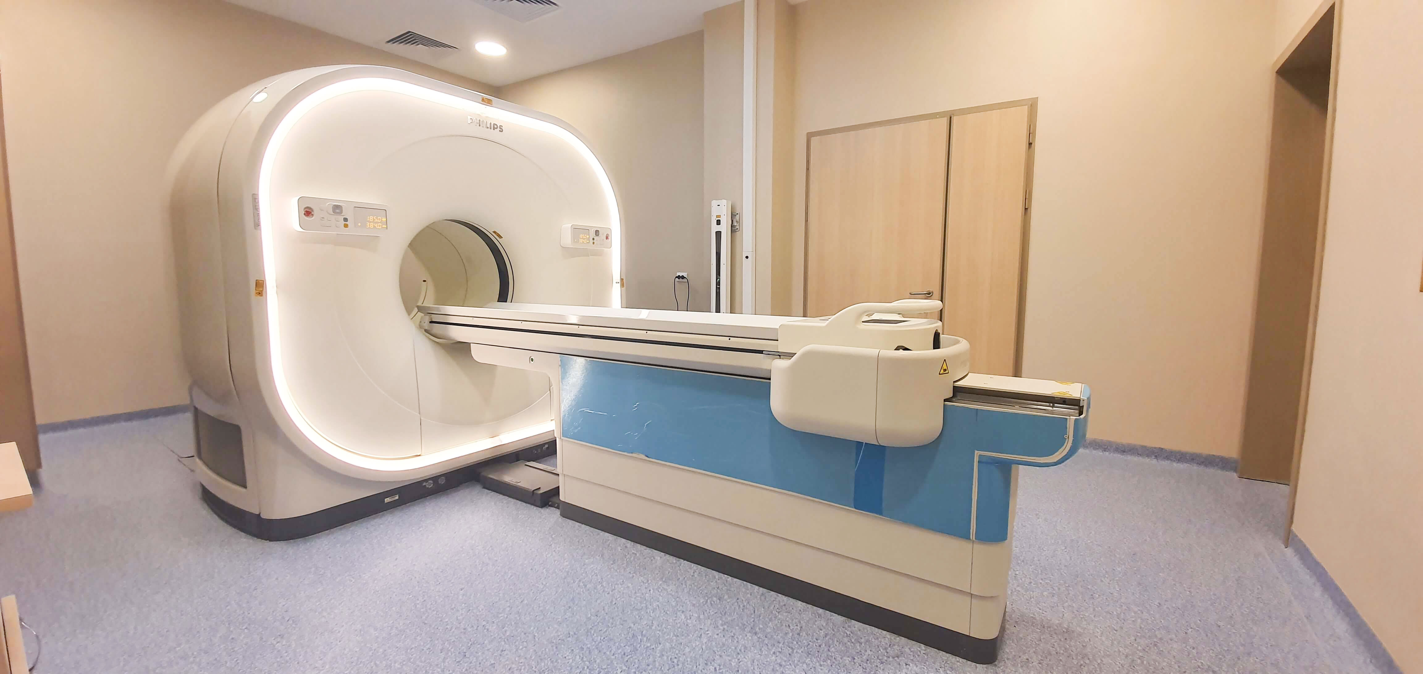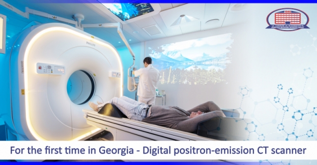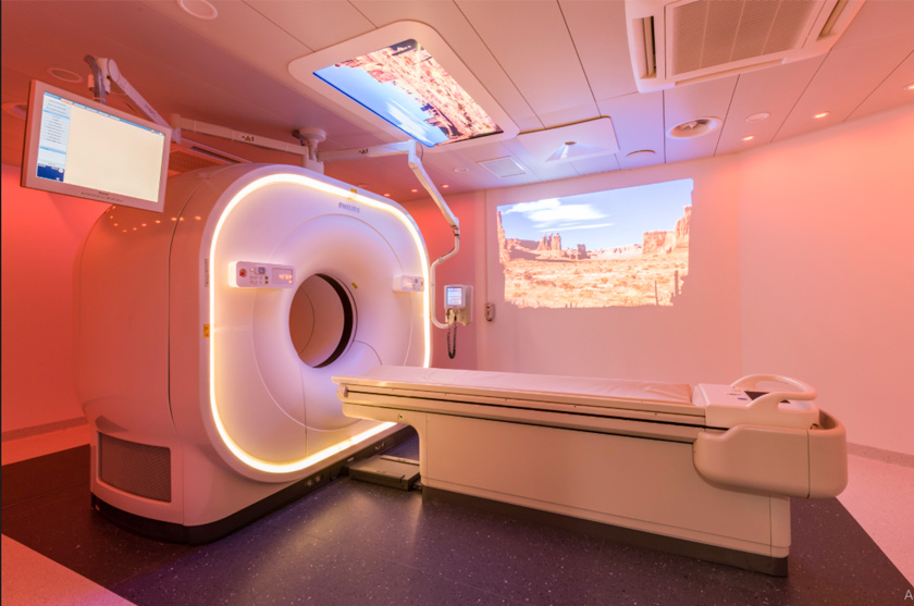
For the first time in Georgia, in Eastern Europe, Asia, and the post-Soviet space, Digital Positron-Emission Computed Tomography – PET / CT was installed at the National Center of Surgery
Philips Vereos Digital PET / CT is the world's first and only fully Digital-Positron Emission Computed Tomography.
The National Center of Surgery gives oncology patients living in Georgia and abroad a unique opportunity, to conduct the highest quality and the most important diagnostic research!
What is Positron Emission Computed Tomography (PET / CT)?
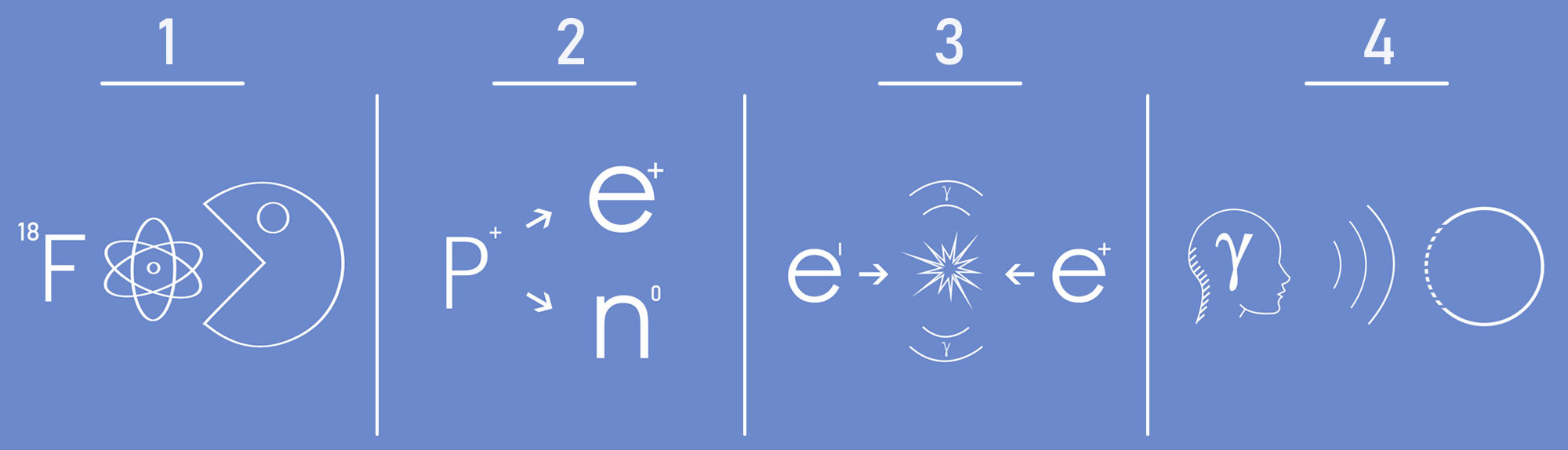
PET / CT is a hybrid study of nuclear medicine. It combines Positron-Emission and Computed Tomography, summarizing the data of which reveals the exact localization of the formation and its metabolism. Tumor cells, compared to healthy tissues, are characterized by enhanced glucose metabolism, while metabolic changes always precede anatomical ones. The research is carried out with the help of a radioactive isotope (FDG - fluorine 18 fluoro-deoxy-glucose), which accumulates in metabolically active cells. A radioactive isotope „marks" glucose in the cell, which is characterized by the emission of positrons (so-called firing) after decay. As a result of the annihilation (interaction) with positron electrons, the generated gamma-radiation is reflected on the detector, which provides information on the current metabolic activity in the cells of the body.
PET/CT can detect tumors that are not visible on computed tomography, during magnetic resonance imaging and other radiological examinations.
That is why Positron-Emission Computed Tomography is a mainstream research method for the early diagnosis and treatment of oncological diseases.
Advantages of digital PET / CT compared to „analog" devices:
- Is distinguished by digital photon counting technology. Registers all the unique photons that exist as a result of isotope decay in the cell;
- High image quality at the expense of increased sensitivity;
- Allows us to accurately describe the formation;Detects even the smallest tumor foci, that do not exceed 2-3 mm and could not be detected by the „analog" device;
- Consumes a lower dose of the isotope, subsequently, digital PET / CT is less sensitive towards its half-collapse and the patient receives less radiation load;
- The research is conducted in a minimum amount of time for the patient.
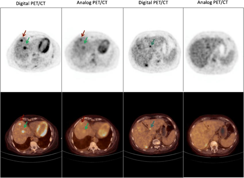
Thus, the benefits of Philips Vereos Digital PET / CT allow us, to detect millimeter-sized tumors, which helps the doctor to diagnose tumors at an early stage and to guide treatment tactics correctly.
PET / CT scan changes treatment tactics in 36% of oncology cases!
In what cases is it necessary to perform Positron-Emission Computed Tomography:
- To get an accurate anatomical and functional picture of the tumor;
- To determine the degree of spread of oncological disease (staging/restoration);
- To identify an unknown primary focus during the metastatic process or paraneoplastic syndrome;
- For the differentiation of benign and malignant tumors;
- To evaluate the effectiveness of the treatment (during oncological surgery, chemotherapy, radiation therapy);
- To select the exact localization of the biopsy of the tumor;
- To plan surgical intervention and radiation therapy;
- For the differentiation of postoperative fibrous tissue and recurrence;
- To determine tumor recurrence or continued growth, when the clinical and anatomical data are negative (e.g., against the background of elevated tumor markers);
- It is necessary for diagnosis both in the first stage and after the treatment and after the end of treatment;
- To evaluate the effectiveness of diagnosis and treatment of lymphomas in hematology.
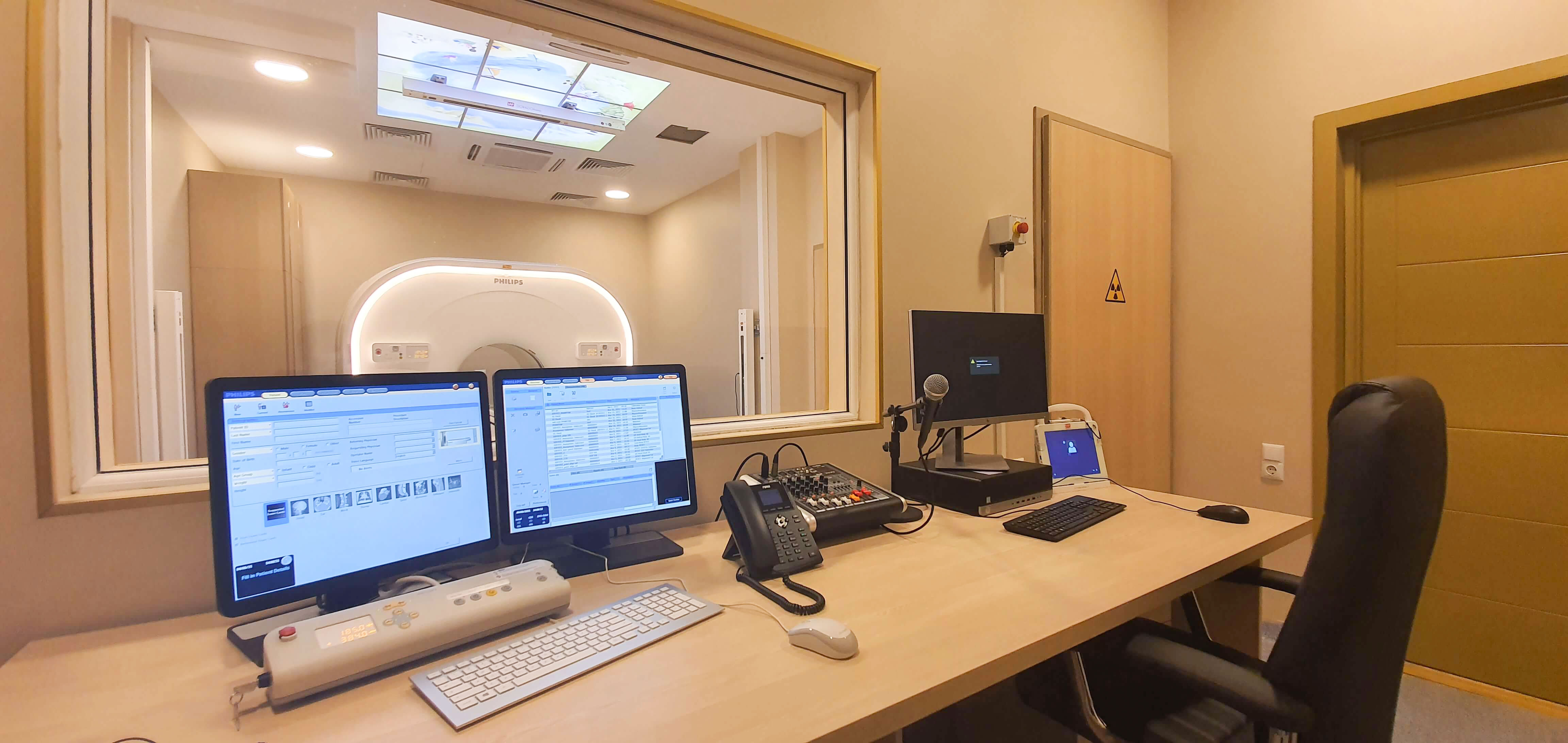
How the procedure is conducted
The patient receives the isotope intravenously. It is necessary to delay one hour from the injection to the shooting. The maximum time for examining the whole body is 20 minutes. The isotope dose is diagnostic only and has no side effects. Isotope distribution in the body is recorded by Positron-Emission Computed Tomography. Most importantly, the patient receives only a minimal amount of radiation in the process of this research.
The Department of Nuclear Medicine complies with international norms in the field of radiation safety.
International Support
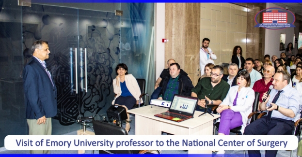
The National Center of Surgery is fully supported by Raghuveer K. Halkar, Director of the Department of Nuclear Medicine, Grady Memorial Hospital, Emory University. He arrived in Georgia in 2019 and presented a report at the National Center of Surgery, which has been devoted to the role of Positron-Emission Computed Tomography in modern oncology. Shorena Esiashvili is a doctor-specialist in nuclear medicine. She began her career in nuclear medicine in 2004 in the Department of Nuclear Medicine, Emory University, the USA, where one of the first Positron Emission Computed Tomography functioned.
The National Center of Surgery offers a full range of services to oncology patients.
The method of treatment of oncology patients in our clinic is determined by a multidisciplinary approach. The case of each patient is treated on the Onco-Concilium, the so-called-Tumor Board, as a result of joint deliberation and discussion.
Together with the clinic physicians, are actively involved in the planning and management of cancer patients, the Head of the Quality Department of Radiation Oncology, Emory University- Professor Natia Esiashvili and the Leading Specialist of the Multidisciplinary Institute of Oncology, „Genesis Care, Spain, Maia Jughashvili-Hernandez, Associate Professor at the University of Murcia, Member of the International Relations Committee of ESTRO International Radiation Oncologists.
Without Positron Emission Computed Tomography, making accurate diagnoses and defining treatment tactics for cancer patients is impossible!
Proven accuracy leads to trust. It is possible to defeat cancer with the National Center of Surgery!
Wish you health!