The radiologists at the International Center of Surgery's Tbilisi and Batumi Clinics will write the conclusions.
The Batumi clinic of The National Center of Surgery offers another important news.
West Georgians are given the chance of getting a comprehensive diagnostic examination of their bodies in Batumi using a digital 3 Tesla magnetic resonance imaging (Magnetom Vida 3T) made by Siemens, a German company, for the first time in the region!
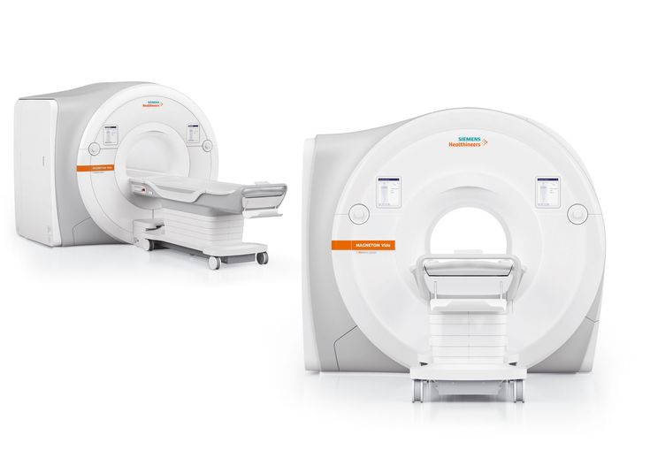
The Siemens Magnetom Vida 3T MRI system features state-of-the-art technology that sets new standards for image quality and diagnostic accuracy.
Radiological service
The clinic established a radiological "board" that adheres to international standards, is staffed by highly qualified personnel provided with modern programs was formed in the clinic, which reviews every study that is completed and provides results.
Leading experts from The National Center of Surgery's Tbilisi and Batumi Clinics will write the conclusions.
With its strong and cutting-edge Tim 4G design, the 3 Tesla MRI produces the best possible images, enabling radiologists to identify even the smallest anatomical structures and ensuring precise diagnosis.
A customized strategy for outstanding outcomes:
The BioMatrix technology of the Siemens Magnetom Vida 3T MRI system takes customization to a whole new level. With customized scanning parameters, it automatically adjusts to each patient's unique anatomical traits. This translates into a quicker and more effective resolution, which eventually enables us to diagnose the patient and provide a personalized treatment plan more quickly.
This unique function minimizes scan time and optimizes image quality by adjusting to the specific anatomy of each patient.
Prioritizing the comfort of the patient
Some patients may find an MRI to be an uncomfortable procedure. The Siemens Magnetom Vida 3T MRI equipment was selected due to its ability to prioritize patient comfort while maintaining picture quality. The patient's peace of mind during the examination is enhanced by its more airy Contour coils and short, light BioMatrix, which guarantee a comfortable examination.
The system incorporates Quiet Suite technology set up which lowers noise levels and produces a peaceful atmosphere for study.
Spectroscopy- ensures an accurate and timely diagnosis
The capability of magnetic resonance imaging to perform spectroscopy (MRS) allows for the analysis of signals produced by different metabolites to ascertain the chemical composition of tissues. It offers insightful knowledge regarding tissue metabolism. mostly utilized for tumor evaluation and neuroradiology.
Due to spectroscopy, in addition to morphological pathology, functional changes can also be detected, allowing for the diagnosis of cellular alterations that take place prior to the formation of pathology. An early diagnosis of the disease is vital for determining the course of treatment.
Benefits and technological features of magnetic resonance imaging in comparison to earlier and comparable devices:
• Magnetom Vida incorporates BioMatrix technology, enabling intelligent patient customisation;
• HyperBand reduces overall scan time. This technology is particularly beneficial for patients who have difficulty staying still;
• Compressed Sensing: is an innovative visualization technique that is revolutionizing the field of medical imaging;
• GOBrain: Magnetom Vida 3T integrates the GOBrain application, which allows a comprehensive neurological examination of the brain in just five minutes;
• Quiet Suite technology aims to reduce acoustic noise during MRI scanning. It makes use of both software and hardware advancements;
• Motion artifacts are minimized and image quality is enhanced by free-breathing imaging;
• Native Direct MR arthrography is a feature of the Magnetom Vida 3T which allows high-quality imagine acquisition without the need for additional contrast agents. It is especially useful for assessing damage to the cartilage and tendons;
• TimTX TrueForm: this technology improves image quality and minimizes artifacts, particularly in challenging clinical situations involving large patients or particular body parts that are difficult to access;
• Functional MRI (fMRI): used to study brain functions. detects changes in blood oxygenation to check brain activity;
• Cardiac MRI: Cardiac MRI allows to evaluate the structure and function of the heart. It provides comprehensive details regarding myocardial viability, blood flow, and heart dimensions.
Batumi Clinic of the National Center of Surgery wishes you health!