Modern medicine is unthinkable without magnetic-resonance tomography.
In radiology, it’s a safe and effective study method for the anatomical and physiological study of the body.
System uses a powerful magnetic field and radio waves for imaging. In the study, human tissue and internal organs aren’t irradiated on an x-ray basis and ionizing radiation isn't used.
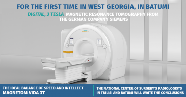
National Center of Surgery is equipped with ultramodern tomography scanner Toshiba Vantage Elan 1.5 Tesla, which is one of the best models in the medical market with its' functions and latest technological packages.
Device is equipped with the following systems to provide a maximally comfortable environment for patients:
- Gentry’s internal lighting that dismisses the notion of closed space;
- MRT bed is a copy of human anatomy and that's why the patient lies comfortably during the study;
- Tomography scanner is equipped with a unique technology called PIANISSIMO, using which the noise is significantly reduced in scanning, study practically progresses without noise and hence, the patient feels comfortable.
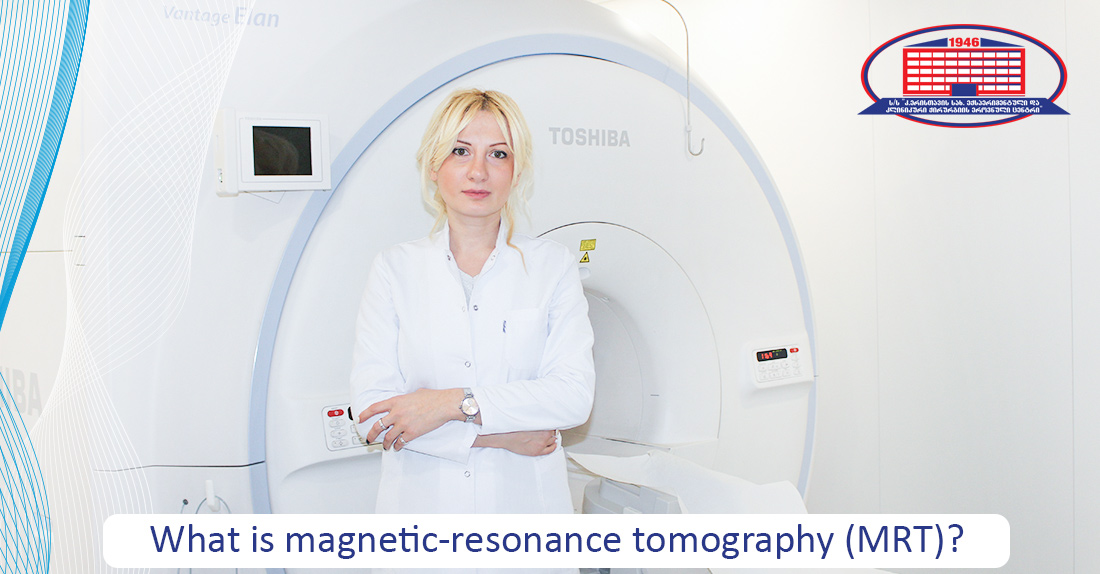
What is magnetic-resonance tomography (MRT)?
Radiologist views any pathological changes proceeding in the body on the screen without cutting into the body. That’s why it's called a gold standard and irreplaceable method necessary for an accurate diagnosis.
MRT study is essential:
- For diagnosing the most acute stage of ischemic stroke;
- It’s informative in demyelinating pathologies;
- In central nervous system diseases;
- In the case of cerebral cortical dysplasia and structural alternation of the spinal cord;
- For diagnosing focal epilepsy;
- In neurovascular conflicts;
- For diagnosing pituitary macro and especially microadenoma;
- In spinal pathologies and so on.
Radiologist of National Center of Surgery Ana Samkharadze discusses the topic.
–What is the difference between magnetic-resonance tomography and computed tomography?
– MRT study is conducted for diagnosing diseases of the brain, spinal cord, pelvic cavity, abdominal organs, soft tissue, joints.
Computed tomography is mostly used for emergency examinations, for example, during traumas, brain hemorrhage, traumatic injuries of various organs. Organs of the thoracic cavity are studied on a planned basis.
It’s worth mentioning that these two studies are simultaneously necessary in some clinical cases and commonly, it’s required in polytrauma, vascular and oncologic diseases!
–What can you tell us about the role of MRT in prompt disease prevention?
– The mentioned study enables the detection of various diseases and pathologies at an early stage, which itself is a precondition for prompt and effective treatment.
In National Center of Surgery, full-body magnetic-resonance screening is ordered to detect metastasis in oncology patients.
Full-body screening is advised on a prophylaxis basis to diagnose a disease at an early stage in clinically undetected pathologies.
It’s recommended for people affected by carcinophobia (fear of developing cancer) and for those who are constantly working because with MRT screening health condition is assessed in the shortest time without staying at the hospital.
It’s completely safe for patients and repeat of the procedure, if necessary, is not limited.
–Is it recommended for a pregnant woman?
– In the first trimester, it's recommended only if it's vital, whereas study is conducted normally from the second-trimester, it doesn't harm the health of a mother and fetus.
– How do you ensure the patient's comfort during the study?
–Each member of the team aims to conduct the study without discomfort. That’s why we offer patients a special pillow, covers, footrest.
Study is conducted with anesthesia for claustrophobic patients.
Wish you health!