The Positron-Emission Computed Tomography at the National Center of Surgery
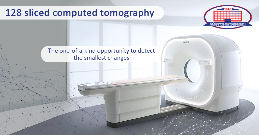
The advantage of a 128-slice Computed Tomography Scan is early disease identification! A
multilayer scanner identifies even the smallest growths, which in itself reveals pathology and
allows us to observe many things, including, possible oncological diseases!
Did you know that David Townsend developed the conceptual model for the world's first
positron emission computed tomography (PET/CT) in 1991 at the University of Geneva?!
During the process, Swiss oncologist Rudi Egel recommended that he would use the empty
space between the revolving jars of bismuth, and add a computed tomography scanner to the
apparatus, this would provide anatomical information to the surgeons. That's how the concept
for the world's first PET/CT scanner emerged!
Due to the tightness of the components installed on the revolving support of the computed
tomography, it quickly became clear, that such a concept would be impossible, however, 7
years later, in 1998, as a result of David Townsend and Professor Ronald Nutt's collaborative
study, The University of Pittsburgh Medical Center now owns the first combined PET/CT
machine!
In 2000, the Positron-Emission Computed Tomography created by the technological
revolutionaries of the medical world gained worldwide recognition and was named the Medical
Invention of the Year!
As an American writer and literary critic Alfred Kazin phrased it: „Art is continually changing, but
it can never evolve like medicine and technology! “
The fact that only 20 years after its initial invention, the PET/CT scanner has improved
technologically to the point where it has switched from analog to digital is proof of this, the
examination duration has been lowered, the scan has become fully safe and comfortable for the
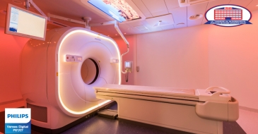
patient, and we can now firmly claim that the Philips Vereos Digital PET/CT at the National
Center of Surgery does not have hidden disorders!


