PHILIPS INGENIA ELITION X
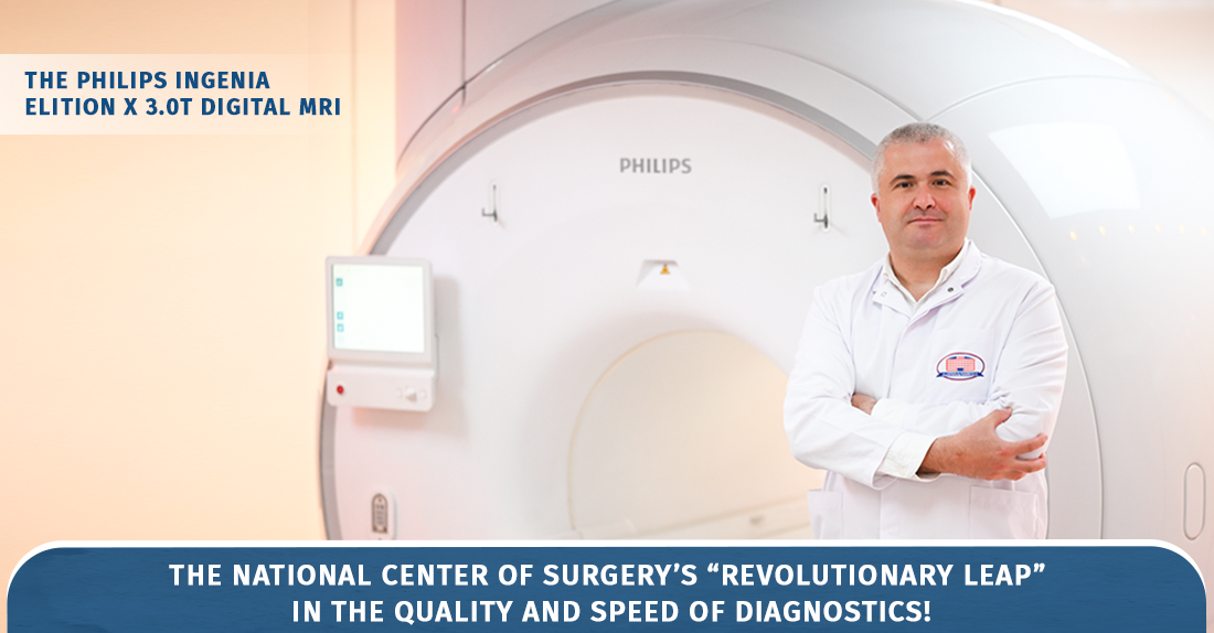
3.0 Tesla Digital MRI PHILIPS INGENIA ELITION X National Center of Surgery's "revolutionary leap" in diagnostic quality and speed!
The capabilities of the new magnetic resonance imaging machine are astonishing
The Konstantine Eristavi National Center for Surgery upholds tradition by keeping the clinic outfitted with the newest, most distinctive equipment available. This time, he introduces a 3 Tesla digital magnetic resonance imaging scanner, one of the most recent medical innovations that is thought to represent a significant improvement in diagnosis speed and quality. What are its benefits and how is it different from earlier analogs? A radiologist named Mr. Tamaz Jaoshvili, Head of the Radiology Department of the Konstantine Eristavi National Center for Experimental Surgery, discusses these and other intriguing innovations.
This time around, the National Center of Surgery also stays true to its heritage by continuing to acquire cutting-edge, distinctive technical equipment. PHILIPS INGENIA ELITION X 3.0T, a 3 Tesla digital magnetic resonance tomography machine, is one of these.
With its features, this cutting-edge 3 Tesla MRI scanner is genuinely unmatched in the Georgian market! One of the most recent medical innovations, the most potent and quick 3-tesla magnetic resonance tomograph (considering its capabilities), was recently given the following motto by the Philips company: "A revolutionary leap in the quality and speed of diagnosis!"
What distinguishes it from current MRI equipment and what are its benefits?
It is important to note that since magnetic resonance imaging does not rely on ionizing or X-ray radiation, it is an entirely safe research technique. All age groups, including second-trimester pregnant women, can utilize it. Because cancer patients frequently require repeated radiological exams, the safety element is particularly crucial for them.
The PHILIPS INGENIA ELITION X, a 3 Tesla digital magnetic resonance imaging scanner, offers extra features and modes, such as COMPRESSED SENSE, which cuts down on examination time by 40–50% without sacrificing image quality.
Instead of a belt, artificial intelligence's VITAL Eye uses lasers to regulate respiration and shields us from motion-induced artifacts, or "poor image quality." For instance, holding your breath during abdominal imaging causes discomfort for the patient, even if we encourage them to do so in order to get the best possible images. VITAL EYE is the very feature that "learns" how often the patient breathes and automatically collects data during phases when breathing is not occurring.
We provide audio-video support for the patient's comfort: the user selects the preferred video-audio file, we create customized glasses for them, and they can watch the video or simply listen to music while the examination is being conducted.
This machine is particularly useful for people who are claustrophobic because it has the biggest tunnel and the highest level of ventilation when compared to other MRI machines.
Patients with "claustrophobia" because the uncomfortable sensation of being in a closed area is reduced by the characteristics and unique lighting field.
It is worth noting separately that, compared to existing devices, the 3 Tesla digital magnetic resonance imaging scanner - PHILIPS INGENIA ELITION X - is designed for a maximum weight (250 kg). It also has COMFORT PLUS equipment - a thick and flexible mattress that alleviates the discomfort caused by lying on the patient's back. There is a widespread belief that during such studies, iron implants are heated and magnetically attracted. In fact, various implants, especially those inserted 10-20 years ago, MRI produces artifacts during the study. Philips took this into account, and our device has the so-called O-MAR reconstruction, through which metal artifacts are reduced and, therefore, implants are no longer a contraindication.
However, which illnesses are the primary targets of magnetic resonance imaging (MRI)?
These days, magnetic resonance imaging is employed in every area of medicine. In this sense, I am only able to draw attention to the locations where our patient flow is especially strong. I refer to neurology and neurosurgery. If magnetic resonance imaging is not employed in the study process, a neurologist and a neurosurgeon are unable to treat any illness.
We can evaluate changes in organ structure, function, and metabolism using the PHILIPS gadget. Regarding the prevalence of herniated discs in the spine, which is rather prevalent in the general population, we need to differentiate between different spine diseases independently. By precisely identifying which intervertebral disc the issue is in and whose neural root it affects, magnetic resonance imaging enables us to choose the best course of action, whether conservative or surgical. Orthopedics and traumatology frequently use these investigations. We can image joints of any size using the equipment and give the doctor a thorough explanation of their architecture.
Make an appointment at your preferred time by calling +995 322 02 25 25
Address: Tbilisi, Dighomi, Chachava №7
Wish you health!
Give us a call