In today's oncology, radiotherapy treatment is critical. Radiation therapy is required for 70% of cancer patients.
Radiation therapy is one of the most effective cancer treatment approaches. So many you know very little about cancer treatment approaches, what the fundamentals of this or that sort of treatment are. The National Center of Surgery's Radiotherapy Department will notify you about the latest radiotherapy treatments.
Radiation therapy has been utilized in medicine for more than a century and is one of the instruments used in cancer treatment. Radiation therapy is absolutely necessary for 70% of cancer patients. Radiotherapy is administered by radiation oncologists to cure the patient or temporarily relieving the illness (palliative treatment).
Radiation Sources
Radiotherapy employs a range of radiation sources, the most common of which is photon radiation. A linear accelerator, X-ray machine, or radiation isotopes accept the photon beam. Additional types of radiation also include particles such as protons and electrons.
Biological Basis And Fractionation Of Radiotherapy
The working principle of radiation therapy is manifested by damage to the cell DNA so that the cell loses its proliferative function. Typically, different types of cancers require a certain total dose of radiation to achieve this effect. Radiation harms the DNA of both cancerous and healthy cells. A cancer cell, in general, has a stronger ability to repair DNA than a healthy one. In addition, cancer cells have a longer radiation-sensitive window in the cell cycle than healthy cells. These two facts also influence the treatment's outcome. In general, the patient is irradiated 5 days per week and given 2 days off during the treatment phase. These two days allow healthy tissues to recuperate. As a result, with the proper dose selection and planning per irradiation, the effect of cancer cell killing is obtained while healthy tissue is preserved. There are several types of tissues that have different dosage tolerances for radiation, these doses are, of course, taken into account while planning treatment.
Clinical Aspects Of Radiotherapy
Radiation therapy kills cancer cells on the tumor's surface. It is given to patients in both radical (healing) and neoadjuvant (preoperative preparation), adjuvant (as supplemental therapy in the postoperative period), and palliative modes (to reduce the symptoms of the tumor and improve the quality of life).
Radiotherapy Is Part Of A Multidisciplinary Approach
Tumors are often treated primarily through surgery, radiation, chemotherapy, or a combination of these methods. Radiotherapy is a crucial aspect of the cancer treatment process in modern oncology for the following localizations: brain, head and neck, lungs, breasts, gastrointestinal tract organs, urogenital system, and numerous metastases of the brain and bones.
Complications Of Radiotherapy
The therapy process can span anywhere from a week to several months on average. During this time, the following difficulties are detectable, which are common for the irradiation of a specific area:
- Skin redness or swelling may develop during breast irradiation;
- Abdominal - nausea, diarrhea;
- Chest - cough, shortness of breath, irritation of the esophagus;
- Head and neck - reduced taste, dry mouth, mucous membrane inflammation, skin redness;
- Brain - hair loss, redness of the scalp;
- Pelvis - diarrhea, convulsions, increased urination, vaginal irritation;
- Prostate - impotence, urinary symptoms, diarrhea;
- Fatigue is a common symptom of large amounts of radiation.
The radiotherapy team works in cooperation to ensure that the patient receives safe and high-quality care.
The team is made up of the following individuals:
- A Radiation oncologist – a physician, who prescribes and administers radiation;
- Medical physicist - guarantees the radiotherapy technique is of high quality. In charge of the calibration and precision of radiation therapy equipment;
- Dosimetrist - creates a radiation plan in collaboration with the radiation oncologist and medical physicist;
- Radiation Technician - conducts the patient's daily procedure to verify that the plan is carried out accurately and with the appropriate dose;
- Radiological Oncology Nurse - communicates with patients and their families during consultations, radiation procedures, and supervision.
Radiotherapy Stages
Consultation, simulation, treatment planning, and quality assurance are the stages of radiotherapy.
Consultation With A Radiation Oncologist
Following a histomorphological confirmed tumor diagnosis, a radiation oncologist will determine whether radiation therapy is appropriate. Taking into account all of the dangers, a treatment plan is developed and coordinated with the full radiotherapy team.
Simulation
The patient is placed on a customized computed tomography to prepare the therapy plan. Fixation devices are used to select the patient's position so that the daily therapy is comfortable and the patient will be able to maintain the same position after the first surgery for the next few days. Healing CT is frequently used for target construction when used in conjunction with magnetic resonance imaging or PET.
Treatment Planning
On the CT scan, the doctor outlines the target volume as well as all the risk organs that are exposed in the radiation field. Medical physicists or dosimetrists create a precise radiation plan using a sophisticated computer program to save as many healthy tissues as possible. The radiation oncologist discusses the treatment strategy.
Safety And Quality Control
During the radiotherapy operation, the greatest emphasis is placed on safety. Each patient's treatment plan is thoroughly examined. The medical physicist ensures that the medical accelerator is accurate and that the radiation dose planned for the patient is correct. A radiation oncologist, dosimetrist, and medical physicist will also review whether a specific plan is being carried out with adequate accuracy daily by a radiation technician.
Radiation Irradiation Procedure
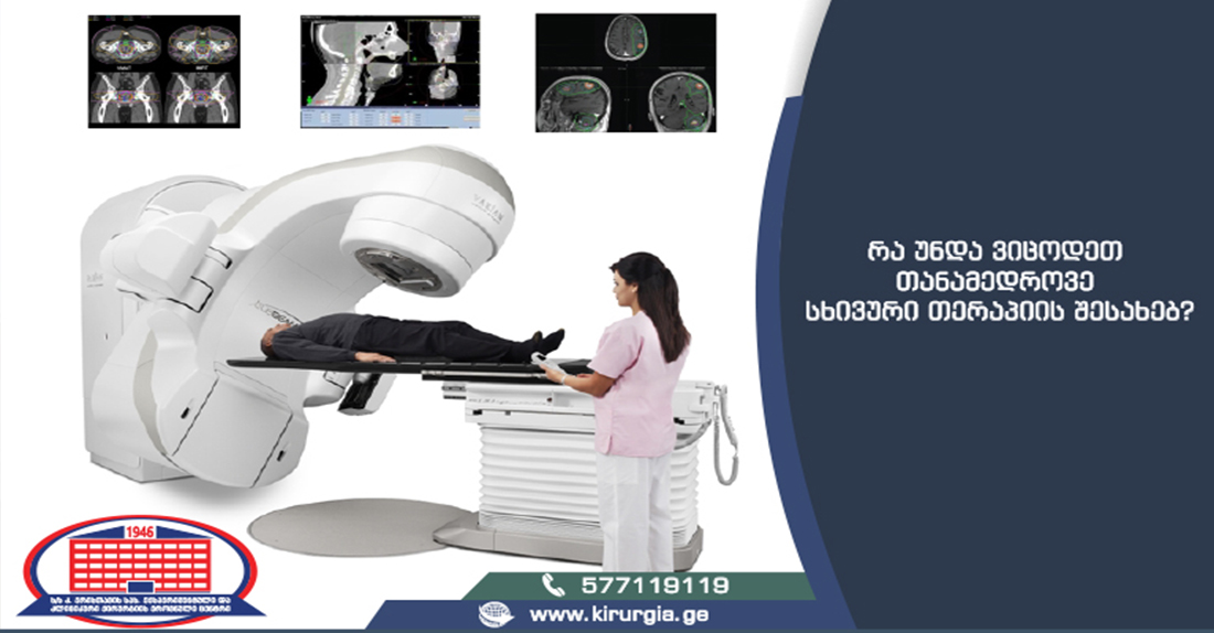
Radiation therapy is classified into two types - remote radiation therapy and invasive radiotherapy. In remote radiotherapy, an external beam is treated via a medical accelerator, but in invasive radiotherapy, the tumor is irradiated directly by inserting a radiation source into the tumor (Brachytherapy, IORT). Gy (Gray) units are used to measure the amount of irradiation.
Remote Beam Radiotherapy
There are different methods of radiation and planning: two-dimensional planning (2D), three-dimensional conformal planning (3D CRT), Intensity Modulated Radiation Therapy (IMRT), Volumetric Modulated Arc Therapy (VMAT RapidArc), Image-Guided Radiation Therapy (IGRT), Stereotactic Radiosurgery (SRS), Stereotactic Radiotherapy (S) Irradiation (Proton, Electron Beams).
Three-Dimensional Conformal Planning (3D CRT)
This sort of planning is frequently utilized in the treatment of breast, breast cancer, palliative care, and low dosages. The dosimetrist decides which fixed directions and how much radiation will occur during 3D CRT. However, collimator sheets shape each field (MLC).
Intensity-Modulated Radiation Therapy (IMRT)
It is a cutting-edge technique for radiation planning in which, the dosage form is customized to the target. This is made possible by collimator sheets, which move in a certain sequence during irradiation to achieve the required dose distribution. The IMRT technique allows us to administer larger doses than the target volume while conserving more healthy tissues, which indicates a higher chance of cure.
Volume Modulated Arc Therapy (VMAT RapidArc)
This is the most recent method of irradiation. The approach employs dynamic MLC, similar to IMRT, in which the field is optimally created. The distinction is that the direction of irradiation is not set, and it is even optimized at the same time. As a result, the dose distribution is the most homogeneous and compliant.
Image-Guided Radiation Therapy (IGRT)
Verifying the patient's position on each fraction of the therapy is part of image management. An X-ray image (KV and MV imaging) of the patient is acquired before irradiation, or a computed tomography (CBCT) scan is conducted with a CT scan. In this approach, the patient can be positioned according to the plan with an accuracy of 1 mm. Regardless of the method of planning, IGRT is now a standard component of remote radiation (3D, IMRT, VMAT RapidArc).
Stereotactic Radiosurgery (SRS)
It is a specific radiation treatment that entails precisely irradiating small, precisely defined targets. This approach is typically used on the brain and is administered in single fractions at massive doses. Of course, this is a tough process that necessitates the highest level of IGRT precision, unique fastening devices, and meticulous planning and execution.
Stereotactic Body Radiation Therapy (SBRT)
It is a procedure similar to SRS in that it involves irradiating areas other than the central nervous system, such as - lungs, prostate, liver, and pancreas. The treatment is typically carried out in big dosages of 1-5 fractions and necessitates excellent accuracy as well as sophisticated fixing devices.
(Brachytherapy)
The formation is irradiated from within by implanting a radioactive source. The major useful sources are 125I, 103Pd, 192Ir, 137Cs. The benefit of brachytherapy is that the radiation penetrates the formation's tissue directly, delivering the dose to the target with the least amount of radiation to the surrounding tissues. High Dose Rate (HDR) brachytherapy is most commonly used for invasive radiation nowadays. It is utilized in gynecological, breast, prostate, and skin irradiation and uses a source of high-energy photons to irradiate the target in minutes.
Intraoperative radiotherapy (Intra Operative RT)
Radiation, like brachytherapy, is an invasive process, with the sole difference being that it is performed surgically after the tumor has been removed. Like brachytherapy, this form of irradiation promotes an effective dose increase, which means greater local control of the tumor and, as a result, an increased possibility of cure. Areas of application are pancreas, abdomen, colorectal, gynecology, kidney, sarcomas, and others.
The National Center of Surgery's Radiotherapy Department will present you with relevant and fascinating material on radiation therapy step by step.
A team of professionals from the National Center of Surgery's Radiotherapy Department is at your disposal!
Wish you health!
What are patients interested in
Chemotherapy
Qauestion:: Hello, My mother received several courses of chemotherapy, because of metastatic involvement of bone and liver. During that time platelet count significantly decreased and only presented with small skin hemorrhages. In chemotherapy, platelet count sometimes increases or decreases, she simultaneously takes Revolade (50mg) on a daily basis and underwent platelet transfusion several times that had an only short-term effect. Is it possible to find another solution for the mentioned problem at your clinic? Thanks in advance