Computed tomography is one of the biggest discoveries of modern medicine.
This is a study that provides the radiologist with detailed information i.e. thorough imaging of organism and enables them to detect various types of pathologies.
Computed tomography is conducted on an elective, as well as urgent basis. It detects muscle and bone pathologies, confirms cancer site, assesses state of viscera, is necessary for cancer monitoring, detects internal traumas or bleeding, etc.
Various surgeries, biopsy and radiation therapy is conducted under control of computed tomography.
The Radiology Department of National Center of Surgery is equipped with ultramodern computed tomography scanner 32-slice Toshiba Aquilion RXL that has innovative study technologies. Perfusion was added to advanced systems of computed tomography scanner of our clinic, with which it’s feasible to study cerebral hemodynamics at the capillary level; This function presents a gold standard in studies and absolute leader in stroke diagnostics.
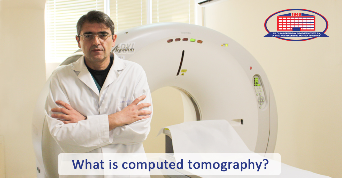
Radiologist of National Center of Surgery, Irakli Gigiadze discusses computed tomography.
– Mr. Irakli, what is computed tomography?
– The term computed tomography is called a procedure (computed X-ray imaging), during which X-ray beams are exposed to the patient. It produces signals processed by computer of the device that generates so-called slices. These slices are called computed images that consist of more detailed information compared to the ones obtained through x-ray exams. After a combination of many similar slices, the computer of the device generates a 3-dimensional image of the patient. This 3-dimensional image provides complete information about primary structures, as well as cancers or other pathologies. Compared to the usual radiograph (that is based on a fixed x-ray tube), a computed tomography scanner uses a motorized x-ray source.
– What type of pathologies is possible to detect with computed tomography?
– This method enables us to study a patient's 3D image (skeletal system, organs, soft tissues). Computed tomography is used for the identification of many diseases and injuries in different parts of the human body. Computed tomography is an effective method for screening of lung and intestine cancers and for diagnosing abdominal cancer. Heart computed tomography is ordered when pathologies of heart and magistral blood vessels are suspected. Head computed tomography is conducted for locating cancer, traumas and for detecting thrombus causing an ischemic infarction, aneurysm, hemorrhage, and other conditions. In the case of chest, it's ordered for diagnosing cancer, pulmonary embolism (thrombus in pulmonary arteries), excess liquid in lungs and also other conditions (emphysema, pneumonia). Computed tomography is essential for assessing complex bone fractures, bone cancer, and joint defect. Computed tomography establishes such life-threating diseases as hemorrhage, thrombosis, cancer. Early diagnostic of these diseases is vital.
– Is it recommended for pregnant women?
– Generally, magnetic resonance imaging or ultrasound is conducted for the examination of the abdominal cavity and lesser pelvis in pregnant women, because with this particular studies the fetus isn't exposed to radiation. Albeit, in cases when listed exams are non-informative or patient’s general health condition requires emergency diagnostics (time is limited) and intervention, computed tomography is perceived to be an acceptable alternative.
– What could we offer for those who are claustrophobic?
– For claustrophobic (fear of closed-off spaces) patients, we explain the purpose of this procedure and how it is conducted. We take them to the operations room and show them how the procedure proceeds for them to overcome the fear. As a trial, we lay them on the CT bed in our presence and imitate the exam by moving the bed. If all of this were without results, we carry out the exam with medical sedation.
– Studies with and without contrast. What does the contrast mean and how are they different?
– Different tissues of the body can block the x-ray beam and because of that, it's quite possible for the image not to be complete for diagnostics. Intravenous contrast is used to solve this problem, which is visible in computed tomography and is safe on an outpatient basis. As an example, iodinated intravenous contrast enhances the assessment of blood vessels and is used for establishing the possibility of vascular obstruction (including heart, urinary system, etc.). Oral contrast is used for diagnosing organs of the gastrointestinal tract.
We don’t use intravenous contrast in case of renal function disorder to avoid permanent failure.
The doctor decides on the necessity of contrast study based on history or obtained tomogram.
Wish you health!